Department of Obstetrics and Gynecology, Faculty of Medicine Siriraj Hospital, Mahidol University, Bangkok 10700, Thailand.
Download:
Abstract
Objective: To assess the sonographic morphology of umbilical cord coiling in chromosomally normal fetuses and estimate the specificity of hypocoiled appearance as a marker for fetal trisomy 21 in the second trimester. Methods: 555 singleton pregnancies undergoing amniocentesis for fetal karyotype were scanned at 17-22 weeks’ gestation. The umbilical cord was evaluated in a longitudinal section using 2D with color Doppler sonography. The cross-sectional data of umbilical coiling index (UCI), defined as the reciprocal of the distance between umbilical coils, were analyzed to establish the normal range according to the gestational age. A UCI less than the 5th percentile was the cut-off value for hypocolied umbilical cord. The specificity of hypocolied umbilical cord to predict fetal trisomy 21 was calculated. Results: As a result of amniocentesis, there were 527 chromosomally normal fetuses and 9 trisomy 21 fetuses. There was no significant difference in maternal characteristics between these two groups. In trisomy 21 fetuses, there was a higher proportion of fetal anomaly indicating for fetal karyotype (22.2% VS 0.6%, p = 0.003). The regression equation of UCI (cm-1, y) on gestational age (weeks, x) was y = 1.205 - 0.033x. The hypocoiled umbilical cord was characterized in 2/9 (22.2%) fetuses with trisomy 21 and in 22/527 (4.2%) chromosomally normal fetuses (p = 0.01), with specificity of 95.83%. Conclusion: The coiling pattern of umbilical cord visualized by sonography has a potential value in second-trimester screening for fetal trisomy 21. Nevertheless, further studies of this model in a larger cohort would provide more information in sensitivity and predictive values.
Keywords: Second trimester, color Doppler, specificity, trisomy 21, umbilical coiling index
Page: 170 - 172
Nowadays increased nuchal translucency (NT) thickness is used as one of the ultrasound markers associated with increased risk of chromosomal abnormalities, especially trisomy 21.1 Approximately 75% of trisomy 21 fetuses have increased NT thickness.2 One of the possible mechanisms of this pathophysiology is the alteration of the extracellular matrix.3 Previous studies had proved the overexpression of collagen VI and hyaluronan in the skin of trisomy 21 fetuses.4-8 Hyaluronan is also the major component of the extracellular matrix in the umbilical cord.9 Therefore the alteration of the amount of hyaluronan in the umbilical cords of trisomy 21 fetuses would affect the morphology of the umbilical cord. The aim of this study is to evaluate the sonographic appearance of umbilical coiling in trisomy 21 fetuses compared with chromosomally normal fetuses in the second trimester and to calculate the specificity of the hypocoiled umbilical cord as a marker for the prediction of trisomy 21.
MATERIALS & METHODS
This prospective study was conducted at the Division of Maternal Fetal Medicine, Department of Obstetrics and Gynaecology, Faculty of Medicine Siriraj Hospital, and approved by the institutional ethics committee (Si 180/2008). Women with singleton pregnancies at 17-22 weeks’ gestation and indicated risk for fetal chromosomal abnormalities were consecutively enrolled in the study from January 2008 to February 2009. Those with chronic diseases or complicated pregnancies were then excluded. Prior to amniocentesis, ultrasonographic examination was routinely performed in detail by physicians with experience in Maternal Fetal Medicine. The gestational age was determined from the certain last menstrual period (LMP). If the discrepancy of gestational age calculated from LMP and fetal biometry was more than 7 days in the first trimester or 14 days in the second trimester, the gestational age was estimated according to fetal biometry.
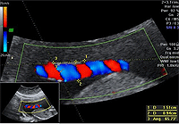
The umbilical cord was then evaluated by three operators, without any information of each patient. 2D-color Doppler ultrasound (RAB 4-8L probe, Voluson E8, GE Medical Systems, Milwaukee, WI, USA) was applied to the free loop of the umbilical cord in the longitudinal view. The image was obtained with maximum magnification so that the discriminatory distance between two points was 0.1 mm. The umbilical vessels and coiling pattern were assessed. The patients were excluded from the study in the case of a single umbilical artery. The intercoil distance was measured from the inner edge of the umbilical artery to the outer edge of the next coil. In each patient, the intercoil distance was averaged from the measurement of two different segments of the umbilical cord (Fig 1). The umbilical coiling index (UCI) was calculated as the reciprocal value of the intercoil distance in centrimetres.7 All the ultrasound findings were recorded in the database after completion of the examination.
Amniocentesis was carried out and fetal karyotype was obtained four weeks later. The presence of fetal chromosomal abnormalities other than trisomy 21 was another exclusion criterion of the study. The nomogram of UCI at 17-22 weeks’ gestation was established. Hypocoiled umbilical cord was defined as UCI less than the 5th percentile for gestational age.
Statistical analysis was performed using SPSS 15.0 software package. The data were presented as percentages or mean and standard deviation. Chi-square and Mann-Whitney U tests were used to compare the qualitative and quantitative data between the two groups, respectively. P < 0.05 was considered statistically significant. Interobserver variability in the measurement of intercoil distance was 0.96.
The relationship between UCI and gestational age was assessed using logistic regression analysis. The normal range and value of UCI at the 5th percentile according to gestational age were estimated. A two by two table was used to calculate the specificity of hypocoiled umbilical cord in prediction of fetal trisomy 21.
RESULTS
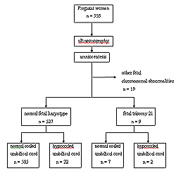
555 pregnant women were recruited in the study after exclusion of cases of chronic diseases, multifetal pregnancies, and single umbilical artery (Fig 2). The result of amniocentesis revealed that there were 9 cases of fetal trisomy 21 and 527 cases of normal fetal karyotype. Meanwhile, there were 19 cases of other chromosomal abnormalities (7 cases of fetal trisomy18, 4 cases of fetal monosomy X, 5 cases of fetal trisomy 13, and 3 cases of fetal triple X), all of which were finally excluded from the study.
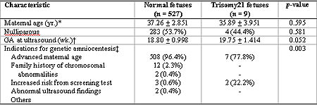
The mean maternal age was 37.2 + 2.9 years and the mean gestational age at ultrasound was 18.8 + 1.0 weeks. There was no significant difference of maternal characteristics between those with normal fetal karyotype and those with fetal trisomy 21, as presented in Table 1. The indications for amniocentesis were advanced maternal age (96.1%), family history of chromosomal abnormalities (2.2%), increased risk from screening test (0.4%), abnormal ultrasound finding (0.9%), and others (0.4%). In 9 cases of fetal trisomy 21, there were 2 cases (22.2%) with abnormal ultrasound findings indicated for amniocentesis, others were indicated due to advanced maternal age.
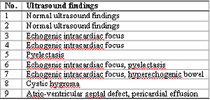
Regarding to the ultrasound findings in 9 cases of fetal trisomy 21 (Table 2), 2 cases (22.2%) had normal ultrasound findings, 5 cases (55.6%) had at least one soft marker of fetal trisomy 21, and 2 cases (22.2%) had at least one major anomaly. In cases of normal fetal karyotype, there were only 3 cases of major anomalies and 1 case with pyelectasis.
The regression equation of UCI (cm-1, y) on gestational age (weeks, x) was y = 1.205-0.033x. In trisomy 21 fetuses, the umbilical coiling index was less than the 5th percentile for gestational age in 2 cases (22.2%). The proportion of hypocoiled umbilical cord between trisomy 21 and chromosomally normal fetuses was significantly different (22.2% VS 4.2%, p = 0.01). Therefore, the specificity of hypocoiled umbilical cord in prediction of fetal trisomy 21 was 95.83%.
Table 1. Maternal characteristics and indications for genetic amniocentesis.
Table 2. Ultrasound findings in cases of fetal trisomy 21.
Figure 1. Measurement of intercoil distance of umbilical cord in 2D color Doppler sonography. Distance 1 represents intercoil distance.
Figure 2. A flow diagram demonstrating the number of participants at each stage of the study.
DISCUSSION
The helical structure of umbilical vessels is fully developed since 8 weeks’ gestation.10 Umbilical arteries have stretched progressively around the umbilical vein and the growth rate of the umbilical cord increases rapidly in the first half of pregnancy. After 24 weeks’ gestation, there is minimal variation of umbilical coiling index due to the slow growth rate of the umbilical cord.11-12 Wharton’s jelly, a connective tissue responsible for the mechanical properties of umbilical cord, is composed of collagens, glycosaminoglycans, proteoglycans, and other microfilaments.9 The over expression of the gene COL1A6 in trisomy 21 fetus has affected the large amount of collagen type VI, contributing to accumulation of hyaluronan.13-14 Superoxide dismutase, encoded by chromosome 21, reduces the degradation of hyaluronan by the mechanism against free radical.15 Hyaluronan has the property of entrapment of a large amount of solvent and inhibits angiogenesis, and therefore has an influence on the growth of umbilical vessels and coiling formation.
The result of the present study revealed that the proportion of hypocoiled umbilical cord in trisomy 21 fetuses is significantly different from that of chromosomally normal fetuses. This finding is observed in the second trimester, which is compatible to the result of the previous study reported in the first trimester7. The high specificity of hypocoiled umbilical cord in prediction of trisomy 21 could be interpreted as the low false positive rate (4.17%). There are a few limitations of this study. Firstly, the study population is the high risk group for fetal trisomy 21, hence the incidence of fetal trisomy 21 is higher than in the normal population and the clinical application of the result is only limited to the high risk population. The small sample size of this study is another limitation that the sensitivity and predictive values could not be calculated. Nevertheless this is the first study demonstrating the sonographic appearance of umbilical coiling in the second trimester of fetal trisomy 21, compared with normal fetuses.
In conclusion, the coiling pattern of umbilical vessels changes accordingly to gestational age. In the second trimester, hypocoiled umbilical cord in trisomy 21 fetuses is more prevalent than in normal fetuses. The sensitivity and predictive values of this model should be calculated from larger studies in the future.
ACKNOWLEDGEMENTS
The authors are grateful to all the physicians who participated in the study, and the nurses who were essential for its completion.
References
1.
Cicero S, Bindra R, Rembouskos G, Spencer K, Nicolaides KH. Integrated ultrasound and biochemical screening for trisomy 21 using fetal nuchal translucency, absent fetal nasal bone, free beta-hCG and PAPP-A at 11 to 14 weeks. Prenat Diagn. 2003 Apr;23(4):306-10.
2.
Bindra R, Heath V, Nicolaides KH. Screening for chromosomal defects by fetal nuchal translucency at 11 to 14 weeks. Clin Obstet Gynecol. 2002 Sep;45(3):661-70.
3.
von Kaisenberg CS, Prols F, Nicolaides KH, Maass N, Meinhold-Heerlein I, Brand-Saberi B. Glycosaminoglycans and proteoglycans in the skin of aneuploid fetuses with increased nuchal translucency. Hum Reprod. 2003 Dec;18(12):2544-61.
4.
Brand-Saberi B, Epperlein HH, Romanos GE, Christ B. Distribution of extracellular matrix components in nuchal skin from fetuses carrying trisomy 18 and trisomy 21. Cell Tissue Res. 1994 Sep;277(3):465-75.
5.
von Kaisenberg CS, Krenn V, Ludwig M, Nicolaides KH, Brand-Saberi B. Morphological classification of nuchal skin in human fetuses with trisomy 21, 18, and 13 at 12-18 weeks and in a trisomy 16 mouse. Anat Embryol (Berl). 1998 Feb;197(2):105-24.
6.
Bohlandt S, von Kaisenberg CS, Wewetzer K, Christ B, Nicolaides KH, Brand-Saberi B. Hyaluronan in the nuchal skin of chromosomally abnormal fetuses. Hum Reprod. 2000 May;15(5):1155-8.
7.
Raio L, Ghezzi F, Cromi A, Cereda E, Passi A. Sonographic morphology and hyaluronan content of umbilical cords of healthy and Down syndrome fetuses in early gestation. Early Hum Dev. 2004 Apr;77(1-2):1-12.
8.
Raio L, Cromi A, Ghezzi F, Passi A, Karousou E, Viola M, et al. Hyaluronan content of Wharton's jelly in healthy and Down syndrome fetuses. Matrix Biol. 2005 Apr;24(2):166-74.
9.
Sobolewski K, Bankowski E, Chyczewski L, Jaworski S. Collagen and glycosaminoglycans of Wharton's jelly. Biol Neonate. 1997;71(1):11-21.
10.
Malpas P, Symonds EM. Observations on the structure of the human umbilical cord. Surg Gynecol Obstet. 1966 Oct;123(4):746-50.
Qin Y, Lau TK, Rogers MS. Second-trimester ultrasonographic assessment of the umbilical coiling index. Ultrasound Obstet Gynecol. 2002 Nov;20(5):458-63.
13.
von Kaisenberg CS, Brand-Saberi B, Christ B, Vallian S, Farzaneh F, Nicolaides KH. Collagen type VI gene expression in the skin of trisomy 21 fetuses. Obstet Gynecol. 1998 Mar;91(3):319-23.
14.
Weil D, Mattei MG, Passage E, N'Guyen VC, Pribula-Conway D, Mann K, et al. Cloning and chromosomal localization of human genes encoding the three chains of type VI collagen. Am J Hum Genet. 1988 Mar;42(3):435-45.
15.
West DC, Kumar S. Hyaluronan and angiogenesis. Ciba Found Symp. 1989;143:187-201.




















 Copyright © 2005-2008 Siriraj Medical Journal