Department of Dermatology, Department of Pathology, Faculty of Medicine Siriraj Hospital, Mahidol University, Bangkok 10700, Thailand.
Download:
Abstract
Eosinophilic fasciitis (EF) is a rare scleroderma-like syndrome. There has been no previous published report of EF in a Thai patient. We described a 41 year-old Thai man who presented with symmetric induration of the skin of forearms, arms, hands, fingers, lower aspects of the legs, and feet. Physical examination revealed bilateral symmetrical woody induration of the skin with peau d’orange appearance. A groove sign was positive on the flexural surface of both arms. Laboratory testing revealed a peripheral eosinophil count of 54%. The skin and superficial fascia biopsy specimen from the inner aspect of the left forearm was consistent with EF. He was treated with prednisolone, methotrexate, and colchicine. He experienced a gradual improvement within 4 months. A history of acute onset of scleroderma-like syndrome and careful physical examination can lead us to the diagnosis of EF.
A 41 year-old Thai man had a one month history of rapid thickening of the skin of the extremities. Thickening of his skin began symmetrically from hands then progressed to proximal arms and legs. He denied fever, night sweating, oral ulceration, photosensitivity, dyspnea, difficult swallowing and Raynaud’s phenomenon. His underlying disease was hypertension. Since three years ago, he has received nifedipine 40 mg/day for hypertension treatment. There was no history of taking any medications or strenuous exercise before the appearance of the symptoms.
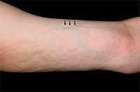
Physical examination revealed bilateral symmetrical woody induration of the skin of forearms, arms, hands, fingers, lower aspects of the legs, and feet with peau d’orange appearance. (Fig 1) A groove sign was positive on the flexor surface of both arms. There were flexion contractures of the elbows, and all fingers with marked decreased passive flexion and extension of the wrist and ankle joints. Muscle strength was normal in the upper and lower extremities. No masked face, sclerodactyly, periungual telangiectasia or Raynaud’s phenomenon were detected. Other physical examinations were unremarkable.
Laboratory testing revealed an initial peripheral eosinophil count of 54% (absolute eosinophil count 10,260 cells/mm3). The erythrocyte sedimentation rate (ESR) was normal. The lactate dehydrogenase level was normal. An antinuclear antibody was positive at the titer of 1:320, speckled pattern. Anti Scl-70 (anti-topoisomerase I) and anti-Sm were negative. Serum protein electrophoresis was normal. Other blood chemistry tests were normal. Chest X-ray and echocardiogram results were normal.
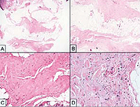
The biopsy specimen from the inner aspect of the left forearm including skin and superficial fascia revealed normal looking epidermis and papillary dermis. The deep reticular dermis and superficial fascia showed marked thickening, fibrosis and sclerotic change with a mixed cellular infiltration composed of lymphocytes, histocytes, plasma cells, and rare eosinophils. (Fig 2) The clinical and histologic findings were considered characteristic of EF. He was treated with prednisolone, methotrexate, and colchicine. He experienced a gradual improvement within 4 months. Absolute eosinophils reduced to 398 cells/mm3.
Figure 1. Induration of skin with peau d' orange appearance and groove sign (arrow).
Figure 2. A&B, Marked thick fibrotic and sclerotic fat septum and fascia (x20 & x40). C, Hyalinization of collagen bundles in the fat suptum and fascia (x100). D, Scattered lymphocytes and plasma cells infiltration in the hyalinized collagen with myxoid change(x400
Table 1. Differential diagnosis of scleroderma-like disorders.6,9
DISCUSSION
Eosinophilic fasciitis (EF) is a rare scleroderma-like syndrome.1 EF was also called Shulman's syndrome or diffuse fasciitis with eosinophilia. It is more common in males.2,3 The mean age of onset is between 40 and 50 years old.1 The etiology is unknown. The followings have been suggested as possibly important as causes of EF; strenuous exercise, initiation of hemodialysis and infection with Borrelia burgdorferi. There are some reports about relationships between EF and some medications such as simvastatin, atorvastatin, fosinopril and dilatin. However, there is no report of nifedipine-induced EF.
The pathogenesis of EF is poorly understood. Some authors suggested an autoimmune phenomenon, supported by increased serum levels of tissue inhibitor of metalloproteinase-1 (TIMP-1) in EF patients rather than healthy individuals. TIMPs are normally found in autoimmune disorders.4 French et al. reported abnormal circulatory clonal T cells and an increased production of cytokine IL-5 which plays an important role regarding production, survival, activation, adhesion and degranulation of eosinophils in EF patients.5
Most patients are characterized by symmetrical induration of the skin with acute onset. Initially, there may be pain, erythema and nonpitting edema of the extremities. The disease progresses to symmetrical induration with orange peel (peau d'orange) appearance. Contracture and rippling of the skin may develop. The most common reported areas of skin involvement include the extremities, neck, and trunk. Some studies demonstrated EF usually spares hands, feet and face.6 However, other studies have shown hand involvement.1,3 Wrist, elbow and ankle joint contractures are commonly present in EF.
When an affected limb is elevated, reducing the distending venous pressure, indentation may be visible along the course of the superficial veins (groove sign). This is due to the relative sparing of the epidermis and superficial dermis by the fibrotic process coupled with the relative immobility of the connective tissue around the remainder of the veins. Thus the superficial layers of skin can bow inward as the peripheral venous pressure falls.
About 60% of patients with EF have peripheral blood eosinophilia during the acute phase of the disease.2 The degree of eosinophilia does not correlate with disease severity and laboratory results are not helpful in following disease activity.1 In addition, ESR is sometimes elevated (29%) and there is often a polyclonal hypergammaglobulinemia (35%).2 Serum levels of creatine kinase are typically normal.7
Magnetic resonance imaging may be used in atypical cases (e.g., fasciitis without skin changes) in order to guide biopsy and monitor the response to therapy. Increased T2 signal and enhancement after gadolinium administration on fat-suppressed T1 images in the subcutaneous and deep fascia have been noted.8
Diagnosis is made by a full thickness skin to muscle biopsy, showing inflammation and thickening of collagen bundles in the superficial muscle fascia with infiltration of lymphocytes and plasma cells, especially in the early period of the disease. Eosinophils are occasionally seen on biopsy, but are not necessary to make the diagnosis.1 In this case, a mild degree of cellular infiltration may be explained from late stage of disease. Differential diagnosis of scleroderma-like disorders has been shown in Table 1.6,9
The commonly used therapy for EF has been corticosteroids, usually equivalent to a prednisone dose of 1 mg/kg per day. Doses are reduced as the affected skin softens. There is usually a rapid resolution of the peripheral blood eosinophilia and normalization of the ESR. If symptoms and signs of EF do not improve and eosinophilia persists, a higher dose of corticosteroids may be necessary.1,6 In a steroid refractory case, cyclosporine, cimetidine, azathioprine, methotrexate, sulfasalazine, d-penicillamine, and psoralen UVA photochemotherapy have all been used successfully for the treatment of EF. Combination of hydroxychloroquine and systemic steroids showed variable results. Topical tacrolimus was used for treatment, but had no effect on the response.1,2
Poor prognostic factors were younger age (under 12 years) at onset, trunk involvement, morphea-like skin sclerosis and prolonged time to diagnosis from the first symptoms.1
References
1.
Bischoff L, Derk CT. Eosinophilic fasciitis: demographics, disease pattern and response to treatment: report of 12 cases and review of the literature. Int J Dermatol. 2008 Jan;47(1):29-35.
2.
Lakhanpal S, Ginsburg WW, Michet CJ, Doyle JA, Moore SB. Eosinophilic fasciitis: clinical spectrum and therapeutic response in 52 cases. Semin Arthritis Rheum.1988 May;17(4):221-31.
3.
Ortega-Loayza AG, Merritt BG, Groben PA, Morrell DS. Eosinophilic fasciitis in a female child. J Am Acad Dermatol. 2008 May;58(5 Suppl 1):S72-4.
4.
Jinnin M, Ihn H, Yamane K, Asano Y, Yazawa N, Tamaki K. Serum levels of tissue inhibitor of metalloproteinase-1 and 2 in patients with eosinophilic fasciitis.Br J Dermatol. 2004 Aug;151(2):407-12.
5.
French LE, Shapiro M, Junkins-Hopkins JM, Wolfe JT, Rook AH. Eosinophilic fasciitis and eosinophilic cellulitis in a patient with abnormal circulating clonal T cells: increased production of interleukin 5 and inhibition by interferon alfa. J Am Acad Dermatol. 2003 Dec;49(6):1170-4.
6.
Connolly M. Systemic Sclerosis (Scleroderma) and related Disorder. In: Bolognia JL, Jorizzo J, Rapini R, eds. Dermatology. 2nd ed. Spain: Mosby; 2008. p. 585-96.
7.
Nakajima H, Fujiwara S, Shinoda K, Ohsawa N. Magnetic resonance imaging and serum aldolase concentration in eosinophilic fasciitis. Intern Med. 1997 Sep;36(9):654-6.
8.
Moulton SJ, Kransdorf MJ, Ginsburg WW, Abril A, Persellin S. Eosinophilic fasciitis: spectrum of MRI findings. Am J Roentgenol. 2005 Mar;184(3):975-8.
9.
Kaufman LD, Krupp LB. Eosinophilia-myalgia syndrome, toxic-oil syndrome, and diffuse fasciitis with eosinophilia. Curr Opin Rheumatol. 1995 Nov;7(6):560-7.





















 Copyright © 2005-2008 Siriraj Medical Journal